The Imaging and Spectroscopy core is primarily located at the Engineering Research Center (ENRC) with a satellite facility available on campus adjacent to the Bioenergetics Core. These facilities currently occupy an area of over 3,000 sq. ft.
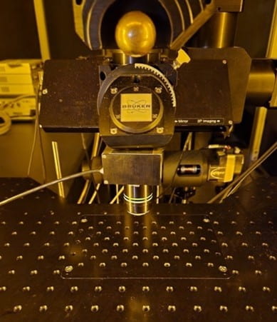
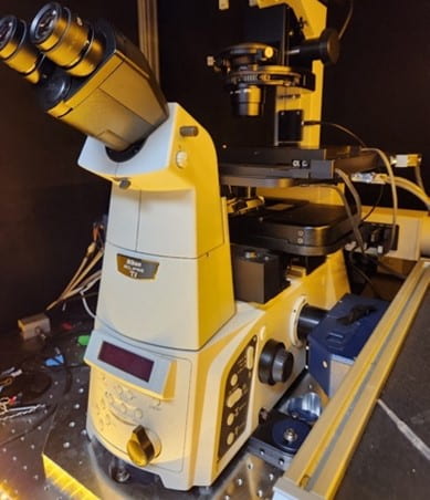
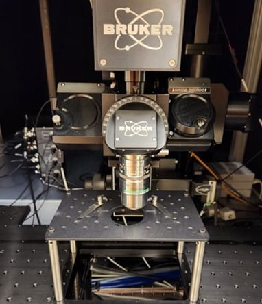
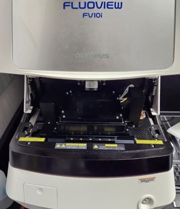
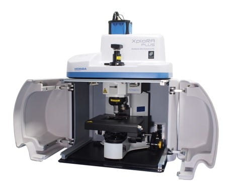
State-of-the-Art Microscopy Equipment Available:
Available Sources of Contrast:
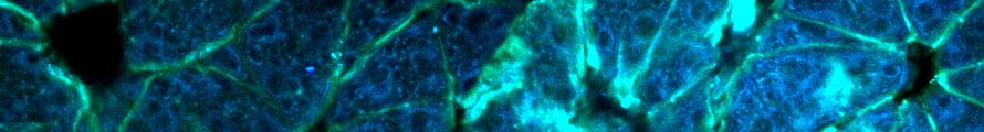
- NADH (460nm emission) and FAD (525nm emission) autofluorescence
- Fluorescence lifetime imaging microscopy (FLIM)
- Coherent anti-Raman scattering (CARS)
- Stimulated Raman scattering (SRS)
Types of Samples:
- In vivo animal studies
- Unstained thin sections
- Thick tissue samples (>1mm)
Available Objective Lenses:
- 20x, 1.0 NA | 2mm working distance | Water immersion
Available Sources of Contrast:
- NADH (460nm emission) and FAD (525nm emission) autofluorescence
- Mitochondrial organization with 60x objective
- Fluorescence lifetime imaging microscopy (FLIM)
Types of Samples:
- Unstained thin sections
- Live cell imaging
- Incubator and 5% CO2 gas mixer available
Available Objective Lenses:
- 60x, 1.2 NA | 0.3mm working distance | Water immersion
- 25x, 1.1 NA | 2mm working distance | Water immersion
Available Sources of Contrast:
- NADH (460nm emission) and FAD (525nm emission) autofluorescence
- Rapid Fluorescence lifetime imaging microscopy (Rapid FLIM)
- Coherent anti-Raman scattering (CARS)
- Stimulated Raman scattering (SRS)
Types of Samples:
- Unstained thin sections
- Live cell imaging
- Incubator and 5% CO2 gas mixer available
- In vivo animal studies
- Thick tissue samples (>1mm)
Available Objective Lenses:
- 25x, 1.1 NA | 2mm working distance | Water immersion
Available Sources of Contrast:
- Exogenous fluorescence (via stains or dyes)
- See “Supported dyes” section below for full list
- Phase contrast
Types of Samples:
- Fluorescently labeled thin sections
- Live cell imaging
- Incubator and 5% CO2 gas mixer available
Available Objective Lenses:
- 10x, 0.4 NA | 3.1mm working distance | Air
- 60x, 1.2 NA | 0.28mm working distance | Water immersion
Available Sources of Contrast:
- Spontaneous Raman spectroscopy
- 532nm, 638nm, and 785nm laser lines
- Single spectra and XYZ mapping
- Brightfield illumination (trans- and epi-direction)
- Darkfield illumination (trans-direction)
Types of Samples:
- Unstained thin sections
- Material characterization
- Live cell imaging *COMING SOON
Available Objective Lenses:
- 5x, 0.1NA | 12mm working distance | Air
- 10x, 0.25NA | 10.6mm working distance | Air
- 50x, 0.5NA | 10.6mm working distance | Air
- 100x, 0.9NA | 0.21mm working distance | Air
Other Free-to-use Equipment:
- Hamamatsu NanoZoomer
- High-speed whole slide imaging system
- Quantitative polarized light imaging (QPLI) microscope
- Capable of quantifying collagen fiber orientation and thickness
- Cryostat
- Handheld diffuse reflectance spectroscopy (DRS) system
- Karl Storz veterinary endoscopy system
- Raman spectrometer
Services:
- Equipment training
- Study design conceptualization
- Data acquisition
- Data analysis for multiphoton microscope images
- Optical redox ratio
- Collagen fiber second harmonic generation (SHG)
- Fiber orientation
- Fiber organization
- Fiber tracking over time
- NADH fluorescence lifetime
- Bi-exponential fitting
- Phasor analysis
- Mitochondrial clustering
- Data visualization and statistical analysis
Please contact Core personnel to schedule access to equipment or discuss experiment plans.
Core Co-Directors

Dr. Narasimhan Rajaram
Associate Professor
Biomedical Engineering
College of Engineering
University of Arkansas
nrajaram@uark.edu
Profile
Dr. Timothy Muldoon
Associate Professor
Biomedical Engineering
College of Engineering
University of Arkansas
tmuldoon@uark.edu
Profile
Core Manager
Dr. Alan Woessner
Research Associate
Biomedical Engineering
College of Engineering
University of Arkansas
aewoessn@uark.edu
Profile