In this post, we’re featuring an exciting UA campus partner that helps us explore our collections inside and out.
Many of the objects in the Museum’s collections stand out for their eye-catching external features, from vibrantly colored bird taxidermy mounts to equally colorful early 20th century crazy quilts.
Vibrantly colored birds and a crazy quilt.
However, valuable information for researchers can also be found within an object’s interior. Enter the MicroCT Imaging Consortium for Research and Outreach. Established in 2018, it is “the home of the University of Arkansas’s micro computed tomography (microCT or µCT) system. MICRO is nested within and managed by the Center for Advanced Spatial Technologies (CAST), which is dedicated to applying geospatial technologies in research, teaching, and service.”
This advanced imaging method is non-destructive and doesn’t require altering the object in any way. When it comes to our collections, MICRO can provide especially great insight into fossils, fluid-preserved zoology specimens, archeological ceramics, and other objects with significant internal structures. It can also provide external details down to the nano-scales (< 1 micron). Learn more about the method’s capabilities here!
Here is a selection of museum objects MICRO has scanned in the last year.
Most recently they were able to scan this telephone from Vol Walker Hall during its previous occupation as the University’s library. The scans allow us to take a look at internal components that allow the phone to work.
Image credit: MICRO and Manon Wilson, MICRO Lab Tech and Senior Research Assistant
This is a Peruvian ceramic from the Museum’s archeology collection. It was one of several ceramics identified as potential whistling pots, which are specifically designed to produce a whistling sound. Learn more about these fascinating vessels from the Peabody Museum! MicroCT scanning was conducted to better understand how its internal structure might enable sound. The images below include both an external and internal scan.
Image credit: MICRO and Manon Wilson, MICRO Lab Tech and Senior Research Assistant
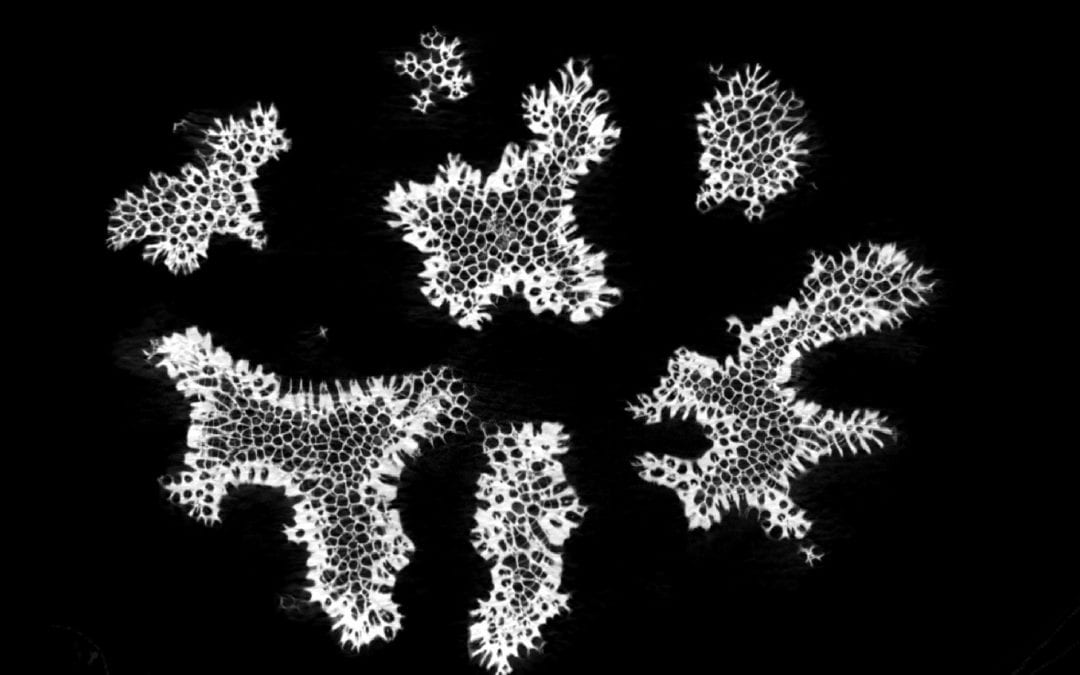
Finally, in the zoology collection, MICRO has provided a highly detailed view of a coral specimen’s complex structure.
Image credit: MICRO and Dr. Claire Terhune, Assistant Professor, Department of Anthropology, University of Arkansas


