Microwear Images
Sample Microwear Texture Images

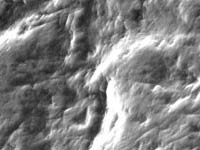
Photomicrograph from a scanning electron microscope (SEM) showing molar microwear on a specimen of Gorilla gorilla. Note the frequent striations and their preferred orientation.
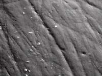
SEM micrograph of heavily pitted molar microwear of Lophocebus albigena. Lophocebus albigena has a diet of harder foods.
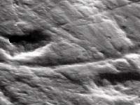
A heavily scratched surface on the second molar of an Oreopithecus bambolii fossil specimen from Tuscany in Italy. This suggests a diet comprised of leaves or other soft objects. (SEM image)
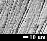
A heavily pitted surface on the second molar of an Ouranopithecus macedoniensis fossil specimen from Macedonia, in northern Greece. This suggests the consumption of harder food items such as nuts or tubers. (SEM image)
Alouatta palliata. Photosimulation based on x,y,z coordinates collected using a white light confocal microscope.

Cebus apella. Photosimulation based on x,y,z coordinates collected using a white light confocal microscope.
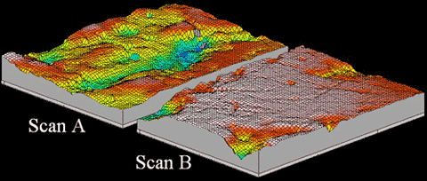
3D meshed axiomatics based on adjacent scans of a white-light confocal microscope. Scans are from molar facet 9 of a specimen of Cebus apella.